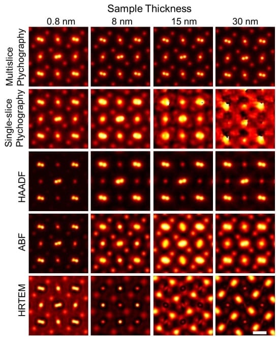
In recent decades, physicists have succeeded in exploring matter on a smaller and smaller scale until it reaches the atomic level. These technologies are necessary to understand the internal structure – hence the mechanics – of the material, but also the technology of the biological tissues. By providing a window on the behavior of atoms in an unprecedented way, the team of researchers has broken a new clarity record thanks to the phytographic algorithm.
In 2018, Cornell researchers built a high-powered discovery that combined with an algorithm-based process called phytography to triple the resolution of an advanced electron microscope, setting a world record. It was successful, and this approach was a weakness. It only worked with samples of a few atoms of dense ultrad. Scatter anything thick Electrons Inseparable.
Recently, a team led by engineering professor David Mல்லller broke their own record with the Pixel Array Detector Electron Microscope (EMPAD) by two factors, which still included 3D reconstruction algorithms. Sophisticated. The resolution is very good, the only dimming left is the thermal agitation of the atoms.
« It didn’t just set a new record. He has hit a diet that is the final limit for resolution. We can now determine where the atoms are in a very simple way. This opens up new possibilities for measuring the matter around us. It also solves a long-standing problem: the cancellation of multiple beam scatterings on the model established by Hans Bethe in 1928, which prevented it from doing so in the past. 2, explains Mller.
Registration resolution Thanks to ptychography
Phytography works by scanning localization patterns from an object model and looking for changes in one another’s region. ” We are looking for speckle patterns that look similar to the ‘laser pointer’ type patterns that attract cats. By looking at how the pattern changes, we can calculate the shape of the object that caused the shape. Says Mller.

To capture the widest possible amount of data, the invention focuses slightly, beam choking. This data is then reconstructed using complex algorithms, resulting in a high-precision image with picometer accuracy.
« With these new algorithms we are now able to fix all the blur in our microscope, the biggest blur factor factor we have left is the atoms shaking themselves because this is what happens to atoms. At a limited temperature 2, explains Mller. ” When we talk about temperature, what we are actually measuring is the average speed of the vibrations of the atoms. .
By using a material made of heavier atoms the researchers can win back their record by making them less oscillating or cooling the sample. But even at zero temperature, the atoms still exhibit quantum fluctuations, so the improvement will not be significant.
Technology with many applications
This latest form of electron phytography allows scientists to detect individual atoms in three dimensions that can be obscured with other imaging methods. Researchers can detect dirty particles in abnormal configurations and capture them one at a time by their vibrations. Imaging semiconductors, catalysts and quantum materials – including those used in quantum computing – as well as materials can be very useful for analyzing atoms in integrated interfaces.
Imaging can be applied to thicker biological cells or tissues or to synaptic connections in the brain – Mல்லller calls them “on-demand connections.” Although this method is time consuming and computationally demanding, it can be developed more efficiently with more powerful computers in conjunction with machine learning and faster inventors.
proof’s: Science

“Avid writer. Subtly charming alcohol fanatic. Total twitter junkie. Coffee enthusiast. Proud gamer. Web aficionado. Music advocate. Zombie lover. Reader.”


More Stories
Acrylic Nails for the Modern Professional: Balancing Style and Practicality
The Majestic Journey of the African Spurred Tortoise: A Guide to Care and Habitat
Choosing Between a Russian and a Greek Tortoise: What You Need to Know